Lung Scan
(Perfusion Lung Scan, Lung Perfusion Scintigraphy, Radionuclide Pulmonary Scan, Ventilation-Perfusion Scan, V/Q Scan)
Procedure overview
What is a lung scan?
A lung scan is a specialized radiology procedure used to examine the lungs to identify certain conditions. A lung scan may also be used to follow the progress of treatment of certain conditions.
A lung scan is a type of nuclear radiology procedure. This means that a tiny amount of a radioactive substance is used during the procedure to assist in the examination of the lungs.
There are 2 types of lung scans: ventilation scans and perfusion scans. A ventilation scan evaluates ventilation, or the movement of air into and out of the bronchi and bronchioles. A perfusion scan evaluates blood flow within the lungs. In the case of suspected pulmonary embolus, both ventilation and perfusion scans are performed either simultaneously or one immediately after the other. If ventilation is normal but perfusion is abnormal, a "mismatch" is said to exist. This mismatch is often indicative of a pulmonary embolus.
The radioactive substance, called a radionuclide (radiopharmaceutical or radioactive tracer), will collect at spots of abnormal blood flow in a perfusion scan. In a ventilation scan, the radionuclide will fill the lungs unless there is an area through which air cannot move. The radiation emitted by the radionuclide is detected by a scanner, which processes the information into a picture of the lungs. Areas in which the radionuclide collects are referred to as "hot spots," while areas of minimal or no collection of radionuclide are called "cold spots."
Lung scans are most often used to diagnose and locate emboli (clots or other small tissue masses) within the blood vessels of the lungs. However, other conditions of the lungs may be evaluated with a lung scan.
Other related procedures that may be used to diagnose problems of the lungs and respiratory tract include bronchoscopy, computed tomography (CT scan) of the chest, chest fluoroscopy, chest X-ray, chest ultrasound, lung biopsy, bronchography, mediastinoscopy, oximetry, peak flow measurement, positron emission tomography (PET) scan, pleural biopsy, pulmonary angiography, pulmonary function tests, and thoracentesis. Please see these procedures for additional information.
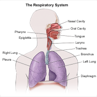
Anatomy of the respiratory system
The respiratory system is made up of the organs involved in the interchanges of gases, and consists of the:

-
Nose
-
Pharynx
-
Larynx
-
Trachea
-
Bronchi
-
Lungs
The upper respiratory tract includes the:
-
Nose
-
Nasal cavity
-
Ethmoid sinus
-
Frontal sinuses
-
Maxillary sinus
-
Sphenoid sinuses
-
Oropharynx
-
Nasopharynx
-
Larynx
-
Trachea
The lower respiratory tract includes the lungs, bronchi, and alveoli.
What are the functions of the lungs?
The lungs take in oxygen, which cells need to live and carry out their normal functions. The lungs also get rid of carbon dioxide, a waste product of the body's cells.
The lungs are a pair of cone-shaped organs made up of spongy, pinkish-gray tissue. They take up most of the space in the chest, or the thorax (the part of the body between the base of the neck and diaphragm).
The lungs are enveloped in a membrane called the pleura.
The lungs are separated from each other by the mediastinum, an area that contains the following:
The right lung has 3 sections, called lobes. The left lung has 2 lobes. When you breathe, the air enters the body through the nose or the mouth. It then travels down the throat through the larynx (voice box) and trachea (windpipe) and goes into the lungs through tubes called main-stem bronchi.
One main-stem bronchus leads to the right lung and one to the left lung. In the lungs, the main-stem bronchi divide into smaller bronchi and then into even smaller tubes called bronchioles. Bronchioles end in tiny air sacs called alveoli.
Reasons for the procedure
A lung scan is most often performed when symptoms, such as tachycardia (fast heart rate), dyspnea (difficulty in breathing), decreased oxygen saturation, chest pain not related to the heart, and other symptoms suggest the presence of a pulmonary embolus.
Other problems of the lungs and respiratory tract that may be evaluated and/or diagnosed with a lung scan include, but are not limited to, the following:
-
Emphysema is a chronic disease in which air spaces in the lungs are enlarged and lose elasticity, or chronic obstructive pulmonary disease (COPD).
-
Tumors or other obstructions in the lungs' blood vessels or airways.
A perfusion lung scan may be performed prior to lung surgery to assess the blood flow and function of the lungs.
There may be other reasons for your doctor to recommend a lung scan.
Risks of the procedure
The amount of the radionuclide injected into your vein or inhaled into your lungs for the procedure is small enough that there is no need for precautions against radioactive exposure. The injection of the radionuclide may cause some slight discomfort. Allergic reactions to the radionuclide are rare, but may occur.
For some patients, having to lie still on the scanning table for the length of the procedure may cause some discomfort or pain.
Patients who are allergic to or sensitive to medications, contrast dyes, or latex should notify their doctor.
If you are pregnant or suspect that you may be pregnant, you should notify your doctor due to the risk of injury to the fetus from a lung scan. If you are lactating, or breastfeeding, you should notify your doctor due to the risk of contaminating breast milk with the radionuclide.
There may be other risks depending on your specific medical condition. Be sure to discuss any concerns with your doctor prior to the procedure.
Certain factors or conditions may interfere with the results of the test. These include, but are not limited to, the following:
-
Remaining radionuclide from a recent nuclear medicine procedure
-
Pneumonia or obstructive lung disease
-
Structural abnormality of the chest
-
Loose or poorly-fitting mask used in a ventilation scan
Before the procedure
-
Your doctor will explain the procedure to you and offer you the opportunity to ask questions that you might have about the procedure.
-
You may be asked to sign a consent form that gives your permission to do the test. Read the form carefully and ask questions if something is not clear.
-
Generally, no prior preparation, such as fasting or sedation, is required prior to a lung scan.
-
Notify the radiologist or technologist if you are allergic to latex or sensitive to medications, contrast dyes, or iodine.
-
If you are pregnant or suspect you may be pregnant, you should notify your doctor.
-
A chest X-ray may be performed prior to the procedure if one has not already been obtained in the previous 24 to 48 hours.
-
Based on your medical condition, your doctor may request other specific preparation.
During the procedure
A lung scan may be performed on an outpatient basis or as part of your stay in a hospital. Procedures may vary depending on your condition and your doctor's practices.
Either a perfusion scan or a ventilation scan or both may be performed. If both types of scans are performed, they will be performed one immediately following the other.
Generally, a lung scan follows this process:
-
You will be asked to remove any clothing, jewelry, or other objects that may interfere with the procedure.
-
If you are asked to remove clothing, you will be given a gown to wear.
-
For a perfusion lung scan, an IV line will be started in the hand or arm for injection of the radionuclide.
-
The radionuclide will be injected slowly into your vein while you are lying flat on the procedure table.
-
After the radionuclide has had time to collect in the blood vessels of the lung, the scanner will begin to take images of the lungs. You will be assisted into several different positions during the procedure so that images of the lungs can be taken from different angles.
-
For a ventilation scan, you will inhale a gas containing the radionuclide through a face mask.
-
After inhaling the radionuclide, you will be asked to hold your breath for a short time. The scanner will begin to take images of the lungs while you are holding your breath. Images will continue to be taken while you breathe in the radionuclide for a few more minutes. Be careful not to swallow the radionuclide, as this could interfere with the images of the lungs.
-
After the radionuclide gas has built up in your lungs, the face mask will be removed. As you continue to breathe normally, the radionuclide will be gradually removed from the lungs.
-
Once the necessary images of the lungs have been obtained, the procedure will be completed. The IV line will be removed.
While the lung scan itself causes no pain, having to remain still for the length of the procedure might cause some discomfort or pain, particularly in the case of a recent injury or invasive procedure such as surgery. The technologist will use all possible comfort measures and complete the procedure as quickly as possible to minimize any discomfort or pain.
After the procedure
You may be monitored in an observation area for a short while after the procedure to assess for any signs of an allergic reaction to the radionuclide.
You should move slowly when getting up from the scanner table to avoid any dizziness or lightheadedness from lying flat for the length of the procedure.
You may be instructed to drink plenty of fluids and empty your bladder frequently for about 24 hours after the procedure to help flush the remaining radionuclide from your body.
The IV site will be checked for any signs of redness or swelling. If you notice any pain, redness, and/or swelling at the IV site after you return home following your procedure, you should notify your doctor as this may indicate an infection or other type of reaction.
You should not have any other radionuclide procedures for the next 24 to 48 hours after your lung scan.
You may resume your usual diet and activities, unless your doctor advises you differently.
For More Information
For more information on how to quit smoking or schedule lung cancer screenings, contact Nancy Sayegh-Rooney, R.N., Pulmonary Nurse Navigator at Richmond University Medical Center, 718-818-2391.
Free screenings are available for at-risk individuals, please call for additional information.